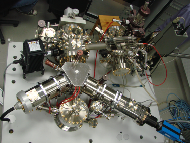
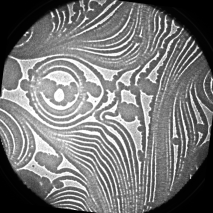
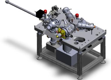
Low Energy Electron Microscope
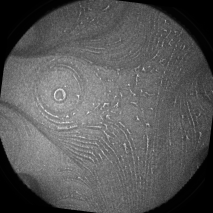
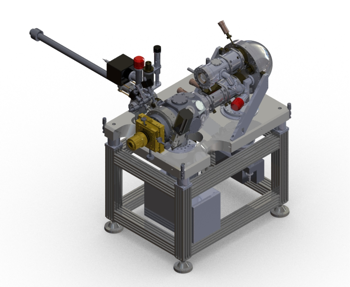
Photoemission Electron Microscope
• field of view: 1.5 - 80μm
• resolution < 3nm

• additional
preparation chamber

Possible extensions
• SP electron gun
• aberration corrector

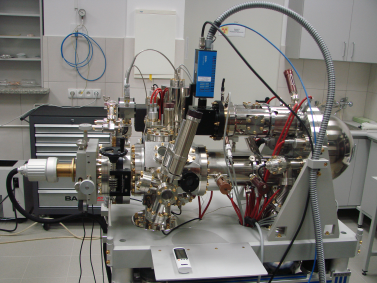
• field of view: 1.25 - 150μm
• resolution < 11nm

• band-pas energy analyser

SPE imaging mode:
• spatial resolution < 10nm
• energy resolution < 0.2 eV
direct imaging energy dispersion:
• energy resolution < 0.2 eV
• directly imaged energy range >15eV

• cooling below 100K
• sample movement around surface normal

meant for Krakow synchrotron
SPE-PEEM at synchrotron enables:
• imaging with
chemical contrast XPS-PEEM, XANES-PEEM
magnetic contrast XMCD-PEEM XMLD-PEEM
• spectroscopies from nanometer scale areas
μ-XPS, μ-XAS